STEP 1 | FOLLOW THE PROCESS
A diagnosis creates chaos.
Understanding replaces chaos with clarity.
Clarity helps you build an effective response plan.
This pathway guides you through what you need to understand and act on, in the order it matters.
Break the diagnosis down into clear steps and take them in the right order.
At each step, understand what matters, why it matters and when it matters.
Earlier right actions help avoid common mistakes and preserve options.
Start The Process
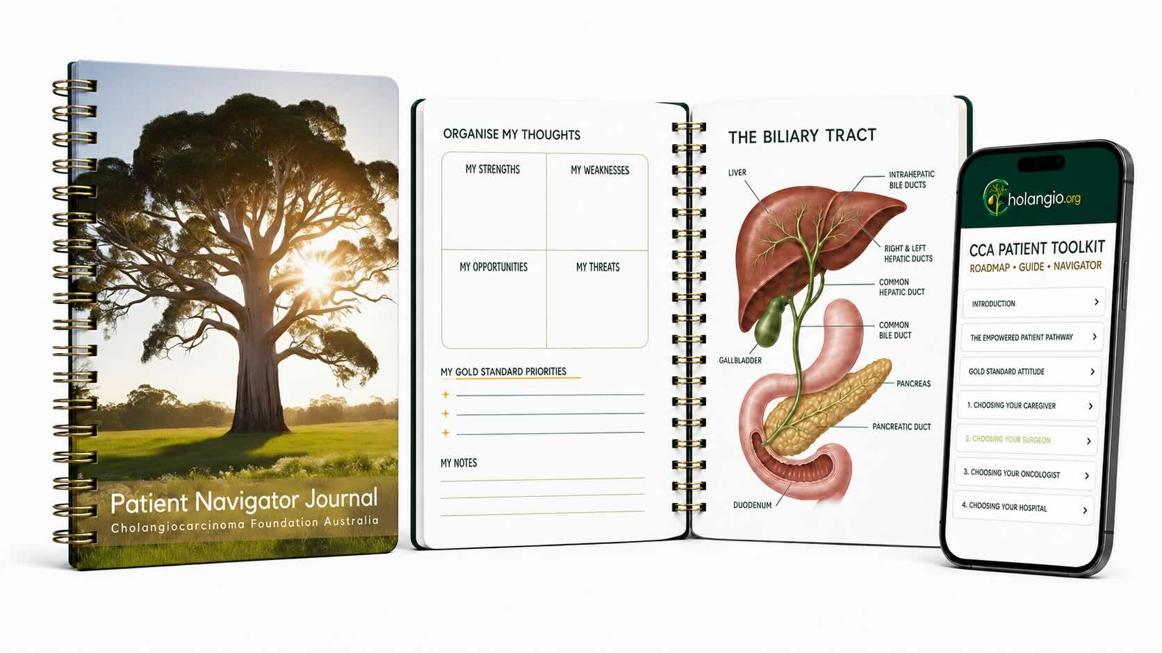
Start with the Patient Navigator Journal
The Patient Navigator Journal puts the process in your hands. It helps you focus on one step at a time, understand it, action it, then move to the next step. That's how we break this cancer down: One Earlier Right Action after the next.
The idea to carry with you
The process is the part you can control.
STEP 2 | PRIMARY TUMOUR LOCATION
Locate your primary cancer.
Where your cancer starts in the biliary system is its primary location. That location helps determine what happens next.
Knowing its location gives you and your doctors a shared starting point for understanding surgery, drainage, biopsy and treatment options.
Visualise it as
The biliary system is a connected drainage network. Your primary location is the point in that network where the cancer began. Where it began changes which structures are involved and which options may be possible.
Understand Your Location
Where did your cancer begin?
Select your primary location to understand what it means. Use the other options when you need them.
Your Next Earlier Right Action
See it. Name it. Record it.
Ask your doctor to show you exactly where the primary cancer started on your scan. Write down its full name and location in your Patient Navigator Journal, then ask what that location changes about the decisions ahead.
STEP 3 | HAS IT SPREAD?
Has the cancer spread?
Knowing where the cancer is now helps determine which treatment options should be pursued and which options need to be protected.
The useful first picture is whether the cancer appears localised, locally advanced or metastatic.
Visualise it as
Your primary tumour is the starting point. Now widen the view. Is the cancer still around where it began, has it grown into nearby territory, or has it established disease somewhere distant?
Understand The Spread
What does your scan show?
Select the description being used for your cancer. Then understand what that description changes.
Your Next Earlier Right Action
See the whole picture.
Ask your doctor to show you the primary tumour and every known site of cancer on your scan. Mark them in your Patient Navigator Journal. Record whether the cancer is being described as localised, locally advanced or metastatic, then ask what that changes about the options you should pursue or protect.
Step 4 | Experienced Medical Opinions
Find the right experience for the decision ahead.
Important decisions about cholangiocarcinoma require specialists with high-volume, hands-on experience in cholangiocarcinoma decisions.
Knowing about cholangiocarcinoma is not the same as regularly diagnosing it, interpreting its pathology, operating on it, selecting treatments or managing its complications.
Each responsibility requires specific expertise.
A specialist may be highly experienced in their medical field but have limited direct experience with cholangiocarcinoma.
What matters is whether the specialist regularly performs the work and makes the particular cholangiocarcinoma decisions affecting your care.
Think of it like
Knowing how to perform a complex task is different from performing it repeatedly.
Experience comes from doing the work, confronting its difficulties and making the decisions it demands.
Match Experience to the Decision
Which decision needs experienced review?
Different decisions require different expertise. Select the decision facing you to understand what experience matters and what you should ask.
Make sure everyone is solving the right problem.
The diagnosis determines what cancer is being treated. Imaging, pathology and other test results must fit together into a clear clinical picture.
Cholangiocarcinoma can be difficult to distinguish from other cancers and conditions. An uncertain diagnosis can lead to the wrong treatment pathway.
A specialist who regularly diagnoses or interprets cholangiocarcinoma brings experience recognising its particular patterns, limitations and diagnostic challenges.
A general cancer diagnosis is not a substitute for the right diagnostic expertise when important uncertainty remains.
The Question
Has my diagnosis been established or reviewed by specialists with high-volume, hands-on experience diagnosing cholangiocarcinoma and interpreting its imaging or pathology?
The decision about surgery requires surgical experience.
Cholangiocarcinoma surgery is highly specialised. Whether an operation is possible depends on the cancer's location, its extent and the technical demands of safely removing it.
A surgeon who regularly performs the relevant cholangiocarcinoma operations brings experience that cannot be replaced by general surgical knowledge.
An oncology opinion, radiology report or multidisciplinary team discussion does not automatically establish that the necessary surgical assessment has occurred.
Step 5 examines this decision in detail, including the opportunity for cure and the importance of direct assessment by an experienced cholangiocarcinoma surgeon.
The Question
Has a surgeon with high-volume, hands-on experience operating on cholangiocarcinoma personally assessed my case?
Treatment choice requires experience treating this cancer.
Cholangiocarcinoma treatment is not simply a choice between chemotherapy, immunotherapy and targeted therapy.
An experienced oncologist must understand the particular disease, its molecular characteristics, the available evidence, clinical trials and the sequence in which treatments may be used.
The objective is to select treatment appropriate to the patient's cancer and circumstances while protecting options that may matter later.
An oncologist who regularly treats cholangiocarcinoma is better positioned to recognise the opportunities and limitations specific to this disease.
The Question
Does my oncologist have high-volume, hands-on experience treating cholangiocarcinoma, including molecularly guided treatment and relevant clinical trials?
Drainage decisions must support the treatment pathway.
Blocked bile ducts can cause jaundice, infection and deterioration in liver function. They can also affect when treatment can begin and which procedures remain possible.
Placing a stent or drain is a technical procedure. Choosing the appropriate drainage strategy requires understanding the particular bile duct anatomy, the obstruction and the treatment being planned.
Experience performing drainage procedures is important. So is experience managing the complex obstructions associated with cholangiocarcinoma.
The Question
Is my bile drainage being planned and performed by specialists with substantial hands-on experience managing cholangiocarcinoma-related bile duct obstruction?
Several opinions do not automatically equal the right expertise.
A multidisciplinary team brings specialists together to examine a patient's diagnosis and treatment options.
That collaboration is valuable, but the presence of different medical specialties does not guarantee that every specialist has high-volume, hands-on cholangiocarcinoma experience.
Nor does a team discussion automatically replace direct assessment by the specialist whose particular expertise is required.
The strength of a multidisciplinary review depends on the experience of the people participating and whether the right expertise is brought to each decision.
The Question
Does my multidisciplinary team include specialists with high-volume, hands-on cholangiocarcinoma experience relevant to the decisions being made, and have those specialists directly assessed my case where necessary?
A second opinion must bring relevant experience, not simply another opinion.
A second opinion is an opportunity to test an important medical decision before acting on it.
Its value comes from the expertise brought to the review.
Another specialist with similar or limited cholangiocarcinoma experience may reach the same conclusion without providing the additional scrutiny needed.
A second opinion from someone with high-volume, hands-on experience in the particular cholangiocarcinoma decision can confirm the recommendation, identify uncertainty or reveal an option not previously considered.
The purpose is not to collect opinions until someone agrees with you. It is to ensure important decisions have been tested by the appropriate expertise.
The Question
Will this second opinion come from a specialist with high-volume, hands-on cholangiocarcinoma experience in the decision I need reviewed?
Your Next Earlier Right Action
Identify the decision. Verify the experience.
Identify the next important decision in your treatment pathway.
Ask which specialist is responsible for making it and whether that person has high-volume, hands-on experience managing cholangiocarcinoma in that particular area.
Establish whether they have personally reviewed the evidence needed to make the decision.
If that experience is absent or uncertain, seek review from an appropriately experienced specialist before an important treatment option is ruled out or an irreversible decision is made.
Need help finding the right experience?
The Cholangiocarcinoma Foundation Australia's Second Opinion Project helps patients identify medical professionals with relevant cholangiocarcinoma experience.
The idea to carry with you
Match the experience to the decision.
Step 5 | Surgery: The only proven pathway to cure
Is there a curative opportunity?
A decision that surgery is not possible today must not close the opportunity for cure without assessment by a surgeon with high-volume cholangiocarcinoma experience.
Cholangiocarcinoma can be cured. Surgery is the only established treatment pathway proven to offer that possibility. Chemotherapy, immunotherapy and targeted therapies may control cancer, extend survival or sometimes make surgery possible, but they are not established standalone cures.
The first decision is whether all known cancer can be completely removed with curative intent, while leaving enough healthy, functioning tissue.
Curative intent is not a guarantee of cure. Even after successful surgery, cholangiocarcinoma can return. But when complete removal is possible, surgery offers an established opportunity that should be assessed early.
Some patients can undergo surgery now. Others may receive treatment that changes what becomes surgically possible later. For some patients, surgery will not become possible.
Liver transplantation is also a surgical curative-intent pathway, available through specialist programs to highly selected patients. It is not a routine alternative to tumour removal.
Think of it like
Surgery is the opportunity to remove the cancer. Drug treatments may slow or shrink it, sometimes enough to make an operation possible. If the door to surgery is closed today, the question is whether it is permanently closed, or whether something could change.
Understand the curative decision
Choose the question that matters to you. One explanation opens at a time.
What does curative intent mean?
Curative intent means treatment is being undertaken with the aim of eliminating the cancer and achieving a cure.
For cholangiocarcinoma, surgery is the only established treatment pathway proven to offer that possibility. The usual surgical goal is to remove all detectable cancer with clear margins, where this can be achieved safely.
A clear margin means no cancer cells are identified at the cut edge of the removed tissue. This is an important surgical goal, but it does not guarantee that cancer will not return.
What about drug treatments?
Chemotherapy, immunotherapy and targeted therapies can have important roles. They may reduce cancer, control its growth, extend survival and, in selected patients, create an opportunity for later surgery.
Some patients experience exceptional, durable complete responses to drug treatment. Those outcomes matter, but they do not establish drug therapy as a reliably curative standalone treatment.
The distinction is between a treatment that may control or eliminate detectable cancer and an established pathway with proven curative potential.
The Question
Is my treatment plan pursuing surgery with curative intent now, trying to create that opportunity later, or primarily controlling the cancer?
Why does surgical experience matter?
Whether cholangiocarcinoma can be removed is both a cancer judgement and a complex technical surgical judgement.
The surgeon must assess the tumour's location, its relationship to major blood vessels and bile ducts, whether it has spread and how much functioning tissue would remain after removal.
Complex operations may require major liver resection, bile duct reconstruction, a Whipple procedure or specialised blood-vessel reconstruction.
Knowing about cholangiocarcinoma is not the same as regularly operating on cholangiocarcinoma.
Radiology reports, oncology opinions and multidisciplinary team reviews are valuable. But a decision that curative surgery is not possible should involve direct assessment by a surgeon with relevant high-volume experience.
That experience strengthens the quality of the decision. It does not guarantee that surgery is possible or that surgery will achieve a cure.
The Question
Has a surgeon with high-volume cholangiocarcinoma experience personally reviewed my imaging and assessed whether complete removal is possible?
Can resectability change?
Resectability describes whether the cancer can be completely and safely removed at the time of assessment.
A cancer may be considered unresectable because of its location, involvement of important blood vessels, spread elsewhere or insufficient healthy liver remaining after surgery.
Unresectable today does not automatically mean unresectable forever.
In selected patients, chemotherapy, immunotherapy or targeted treatment may reduce or control disease enough to justify another surgical assessment. These treatments are not themselves established standalone cures.
In some cases, specialist procedures can help prepare the liver for a major operation. The amount of functioning liver expected to remain is called the future liver remnant.
Not every barrier can be changed. Reassessment should be guided by meaningful changes in the cancer, the patient's condition or the surgical options available.
The Question
What specifically prevents surgery now, could that barrier change, and what would trigger another surgical assessment?
Could transplantation be an option?
Liver transplantation is another surgical pathway with curative intent for highly selected patients with cholangiocarcinoma.
Instead of removing only the affected part of the liver and bile ducts, transplantation replaces the liver as part of a carefully controlled treatment protocol.
It is not a routine alternative whenever conventional surgery is considered impossible.
Eligibility depends on the cancer's location and extent, previous treatments and the entry criteria of a specialist transplant program.
Recognised protocols are particularly relevant to certain carefully selected cases of perihilar cholangiocarcinoma. Other transplant approaches remain restricted to specialist programs or research settings.
Transplantation therefore sits within the broader surgical pathway to cure. It does not make chemotherapy, immunotherapy or targeted therapy established standalone cures.
The Question
Does my diagnosis meet the criteria for assessment by a recognised cholangiocarcinoma transplant program?
Preparing for a surgical second opinion
A surgical second opinion should answer a specific question: Is there a curative surgical opportunity that has not yet been fully assessed?
The surgeon needs the actual scan images, not just the written reports, along with the relevant clinical history and treatment information.
Records to request
- Current CT and MRI images and reports, plus other imaging where relevant.
- Pathology, biopsy and previous operative reports.
- Recent blood tests, including liver function.
- Previous treatments and the current treatment plan.
- Previous surgical and multidisciplinary recommendations.
Take your Patient Navigator Journal. Ask the surgeon to show you the cancer on the images, explain the barriers to surgery and identify what happens next.
Explore the Foundation's second-opinion resources
The Question
Can my imaging and clinical records be sent now to a surgeon with high-volume cholangiocarcinoma experience for direct review?
Questions to ask about cure
The purpose of these questions is to establish whether a curative surgical opportunity exists now, might become possible later, or has been ruled out after appropriate expert assessment.
- Can all known cancer be completely removed with curative intent?
- Has a surgeon with high-volume cholangiocarcinoma experience personally reviewed my imaging?
- What specifically prevents surgery if it is not possible now?
- Could treatment or a specialist procedure change that barrier?
- If drug treatment is proposed, is its goal cancer control, preparation for possible surgery, or something else?
- Is there a recognised transplant pathway that should be considered?
- What would trigger reassessment of the surgical opportunity?
- If surgery is not possible, have biopsy, molecular profiling and other treatment opportunities been addressed promptly?
The One Question
Is there a proven surgical pathway to cure available to me now, and if not, what would need to change for that possibility to be reconsidered?
The Question
Has a surgeon with high-volume cholangiocarcinoma experience personally reviewed my imaging and explained whether a curative surgical opportunity exists now, or could become possible later?
Your Next Earlier Right Action
Secure an experienced surgical assessment.
Surgery is the established pathway offering a possibility of cure. Protect that opportunity by obtaining the right assessment before accepting that it is unavailable.
The idea to carry with you
A door closed to cure today is not necessarily closed tomorrow.
Step 6 | Biopsy the tumour
Tissue is evidence. Protect the curative pathway first.
Before any biopsy route is chosen, every remaining curative opportunity must be protected.
When curative surgery cannot be performed at this time, evidence of what is driving tumour growth becomes the next immediate priority. Obtaining a biopsy of the tumour tissue is how this evidence is secured.
The sample must confirm the diagnosis and leave enough material for biomarker testing and comprehensive genomic profiling.
But the route used to obtain that sample matters. For some patients, particularly those with perihilar cholangiocarcinoma, the wrong biopsy route can affect a possible transplant pathway.
Think of it like
A biopsy gathers evidence needed to understand the cancer. But gathering that evidence must not unnecessarily close a remaining opportunity for cure.
Understand the biopsy decision
Choose the question that matters to you. One explanation opens at a time.
Hilar tumours
An inoperable perihilar tumour may still have a transplant pathway. Before any biopsy route is chosen, it should be reviewed by a high-volume hepatobiliary and transplant team.
Is seeding a risk?
All biopsy procedures carry the risk of seeding. On balance this risk is accepted, but not in all settings. Potential seeding risk for hilar tumours will rule a patient ineligibile for future transplant opportunities.
Obtain enough tissue
The sample must confirm the diagnosis and leave enough material for biomarker testing and comprehensive genomic profiling.
Testing the tissue
Pathology identifies the cancer. IHC biomarker testing reads important signals. Genomic profiling searches deeper for drivers that may open more treatment options.
Why tissue matters
A biopsy is not just a test
A biopsy collects tumour tissue for the pathologist to examine. It can confirm the diagnosis and provide material for the next layers of testing.
Think of the tumour as a damaged city. Histopathology looks at the buildings and identifies what kind of cancer it is. Biomarker testing checks important signals on its surface. Genomic profiling reads deeper into the faulty instructions that may be driving its growth.
One tissue sample can help answer three different questions: What is it? What signals does it carry? What may be driving it?
The Question
Do we already have enough suitable tissue for histopathology, biomarker testing and comprehensive genomic profiling? If not, what exactly is missing?
Before any biopsy, protect surgery and transplant options
Not every biopsy route is interchangeable
A suspected hilar cholangiocarcinoma sits at an important intersection. A person may not be suitable for conventional surgery, yet still have a possible transplant pathway.
In this setting, some biopsy routes through the abdomen or duodenum may create concern about tumour cells being carried outside the bile-duct area. That can affect eligibility for some transplant protocols.
This does not mean tissue is unimportant. It means the pathway must be reviewed before the route is chosen. In selected cases, imaging, bile-duct sampling and other clinical evidence may help establish the diagnosis while the transplant question is assessed.
Bring the right team into the decision
If the tumour is hilar, potentially resectable or potentially transplantable, ask whether an experienced hepatobiliary surgeon or transplant centre should review the imaging and biopsy plan first.
The Question
Could this biopsy route affect surgery or transplant eligibility, and has an experienced hepatobiliary or transplant team reviewed the plan?
How tissue may be obtained
There is no single best biopsy. The safest and most useful route depends on the cancer's location, whether bile drainage is needed, the amount of tissue required and the treatment options that still need protecting.
ERCP or PTC-guided sampling
Where the cancer is in or near the bile ducts, tissue may be obtained from within the biliary system during ERCP or a percutaneous transhepatic procedure. These procedures may also be used to manage bile drainage.
Image-guided or EUS-guided sampling
An interventional radiologist or endoscopic ultrasound specialist may be able to sample an accessible lesion or lymph node. The target and route must fit the individual anatomy and the future treatment plan.
Blood-based testing may add information
ctDNA, sometimes called a liquid biopsy, may detect an important cancer change in the bloodstream. It can add useful information, but a result with no finding does not prove that no relevant change is present.
The Question
Which route protects the most future options while giving us enough useful tissue for the testing that follows?
When tissue is difficult to obtain
Find out what is preventing a suitable sample
A biopsy may be difficult because of tumour location, bleeding or infection risk, limited access, or because an important treatment option still needs protecting. That does not mean the question has been answered.
Ask what is preventing a suitable sample, which specialist has assessed the route, and when the tissue question will be reviewed again.
The Question
What is preventing a biopsy now, who has assessed the safest route, and what plan protects the most options while we obtain the evidence needed?
The Question
Could the proposed biopsy route affect a remaining curative option, and will the sample provide enough evidence for diagnosis and molecular testing?
Your Next Earlier Right Action
Before tissue is taken, protect the best remaining option.
Ask whether the proposed biopsy route could affect surgery or transplant eligibility. Then ask whether it will provide enough tissue for diagnosis, biomarkers and comprehensive genomic profiling.
The idea to carry with you
Protect the pathway before taking the tissue.
Step 7 | Biomarkers Provide Clues
Know your biomarkers.
Your pathology report must establish what the cancer is, reveal the biomarkers that matter and identify what still needs testing before treatment options are lost.
Your initial pathology report, which should include immunohistochemistry (IHC), provides important clues about your diagnosis.
It helps confirm where the cancer began, influence treatment direction including whether immunotherapy or targeted treatment may be an option for you.
Think of it like
An evidence file. The pathology report gathers the clues that help establish where the cancer began, what it is doing and what needs to be tested next.
Understand your pathology evidence
Select a topic to explore its meaning, the evidence it provides and what to ask.
Sufficient tissue
Make sure your doctors know you need enough tissue for pathology and future molecular profiling.
What is missing?
If an item is not recorded, ask your doctor to request it. Not recorded does not mean it is not present.
Request a copy by email
Keep a digital copy of your pathology and IHC report so you can easily share it when seeking another opinion.
What should be looked for in the pathology report?
A pathology report should do two jobs. First, it should show why the cancer is believed to be cholangiocarcinoma rather than a cancer that started somewhere else. Second, it should show whether there are findings that could affect treatment decisions.
Think of the report as an evidence file. It gathers the clues that help establish where the cancer began, what it is doing and what needs to be tested next.
What to look for
- A clear diagnosis and description of the tumour.
- Diagnostic markers that support bile duct origin.
- Markers that may reveal treatment opportunities.
- Tumour differentiation, describing how abnormal the cells look.
- Whether enough tissue remains for comprehensive genomic profiling.
Ask
Can you show me what in this report supports the diagnosis, what has already been tested, what is missing and whether there is enough tissue left for the next tests?
Diagnostic markers that support bile duct origin
Cell Origin
Immunohistochemistry, or IHC
Visualise IHC as a laboratory scanner reading the ID tags carried by cancer cells. Special stains are placed on the biopsy tissue to show which proteins the cells carry.
No single tag proves where a cancer began. The pathologist reads the complete tag pattern, alongside the scan and the appearance of the cells, to build the strongest answer.
Cell Origin
CK7
Think of CK7 as a cell ID tag often carried by bile duct cells. A positive CK7 result can support a bile duct pattern, but it can also appear in other tissues. It is one clue within the full pathology picture.
Cell Origin
CK19
Think of CK19 as a second duct-cell ID tag. It is commonly found in cells with bile duct features. When CK7 and CK19 appear together in the right clinical setting, they can strengthen the evidence for a bile duct origin.
Comparison Marker
CK20
Think of CK20 as a comparison marker that may help identify intestinal differentiation. It is interpreted alongside CK7, CK19 and other markers when assessing where a cancer may have begun. CK20 alone cannot establish or exclude a bile duct origin.
Comparison Marker
CDX2
Think of CDX2 as a directional signpost towards intestinal tissue. It helps test whether the cancer could have begun in the bowel. It is read alongside the other markers, not in isolation.
Comparison Marker
SATB2
Think of SATB2 as a stronger signpost towards the lower bowel. It is often used when the team needs to consider or rule out colorectal cancer as the original source.
Comparison Marker
TTF-1
Think of TTF-1 as a signpost that may point towards lung or thyroid tissue. It can help check whether the cancer may have begun in one of those organs rather than the bile ducts.
Comparison Marker
PAX8
Think of PAX8 as a signpost for kidney, thyroid or some gynaecological tissues. It helps test whether those possible origins should be considered or excluded.
Comparison Marker
GATA3
Think of GATA3 as a signpost that can point towards breast or urinary tract tissue. It may be used when the team needs to distinguish cholangiocarcinoma from cancer that began in those areas.
Ask
Which markers support bile duct origin in my case, and which other possible origins have been considered and ruled out?
Treatment-opportunity markers to request and confirm
Think of these as option-finding signals. They do not guarantee a treatment. They can show that a treatment discussion or another test should happen earlier.
DNA Repair
MMR status
Visualise a cell as a printing room copying its instruction manual before it divides. MMR is the proof-reading team checking every copied page.
MMR means mismatch repair. Its normal job is to find and repair copying mistakes before they become part of the next cell.
Visualise MLH1, PMS2, MSH2 and MSH6 as the four named proofreaders in that team. The report checks whether these proofreaders are present and working.
If the proofreaders are working, the result is usually pMMR, meaning proficient mismatch repair. If one or more proofreaders are missing or not working properly, the result may be dMMR, meaning deficient mismatch repair.
Why it matters: without proof-reading, copying mistakes can build up. This may make the tumour more visible to the immune system and should trigger an early immunotherapy discussion.
DNA Repair
MSI status
Visualise MSI as the trail of typos left behind when the proof-reading team fails. MSI means microsatellite instability. It measures whether copying errors have built up in repeated sections of DNA.
An MSI-high result means many errors have built up. It can be linked to dMMR and may be relevant to treatment options, particularly immunotherapy discussions.
Immune Signal
PD-L1
Visualise PD-L1 as a false stop sign displayed by a cancer cell to slow an immune attack. It can interact with immune cells and reduce their ability to attack the cancer.
A PD-L1 result may help inform an immunotherapy discussion, but it is only one part of the full picture. A low or negative result does not automatically mean immunotherapy is not relevant.
Growth Signal
HER2
Visualise HER2 as a growth antenna on the outside of a cell. It receives growth messages. Too much HER2 signalling can help drive cancer growth.
HER2 is commonly reported on a scale from 0 to 3+. A result of 2+ may need another test to clarify whether HER2 is genuinely driving the cancer and whether a HER2-targeted option should be explored.
Cell Appearance
Tumour differentiation
Visualise differentiation as how recognisable the original cell remains after cancer has changed it. Well-differentiated cells still look more like the tissue they came from and tend to grow and spread more slowly. Poorly differentiated cells look less like normal tissue and tend to grow and spread faster.
This helps describe the tumour's biology and likely behaviour, but does not determine treatment on its own.
Ask
Please confirm whether MMR, MSI, PD-L1, HER2 and tumour differentiation have been tested or reported. If something is missing, can it be requested now?
Why missing markers cost valuable time
Missing information does not always mean a test was negative. It may mean the test was not ordered, there was not enough tissue, the result is still pending or the report does not clearly state it.
Think of this as an incomplete map. The cancer does not wait while information is collected later. If important tests are missing, the team may need to find more tissue, repeat a biopsy or wait for another report before the full range of options can be seen.
Why this matters now
- It may avoid a delayed or repeated biopsy.
- It preserves time for comprehensive genomic profiling.
- It helps treatment decisions begin with the fullest possible picture.
- It reduces the risk that an opportunity is found only after treatment has already started.
Ask
Can you show me which markers have been tested, which are still missing, whether results are pending and whether enough tissue is available to complete the missing tests?
If tissue is not enough or a biopsy is difficult
Do not let "difficult biopsy" end the question. Ask what is preventing a suitable sample, who has assessed the route and what happens next.
First ask whether the tissue already collected is enough for the diagnosis and molecular profiling. If it is not, a biopsy may be needed to obtain more tissue.
The safest and most useful biopsy route depends on where the cancer is, what needs to be sampled and whether surgery or a transplant pathway is still being considered. The procedure should be assessed by the specialist with the right experience for that route.
Blood-Based Testing
ctDNA, also called liquid biopsy
Think of ctDNA as small fragments of the cancer's changed instruction plans travelling in the bloodstream. A blood test may find a treatment-relevant change while the tissue question is being assessed.
A blood test can be useful, but it does not replace tissue in every situation. Some cancers do not release enough tumour DNA into the blood to be detected. A blood result with no finding does not prove that no relevant change is present.
Ask
Is existing tissue enough for molecular profiling? If not, what is preventing a biopsy, has the right procedural specialist assessed the safest route, is ctDNA testing appropriate now and when will the tissue question be reviewed?
The Question
What does my pathology report confirm, which biomarkers have been identified, and what is still missing before my treatment decisions are made?
Your Next Earlier Right Action
Protect the evidence from the beginning.
Obtaining a tissue biopsy and finding the tumour's biomarkers is vital. For some people, getting enough tissue and the information it holds will require persistence.
The idea to carry with you
Find the clues before options are lost.
Step 8 | Genomics dive deeper
Understand what is driving the cancer.
A genomic result becomes valuable when it identifies what is driving the cancer and changes a treatment decision before an opportunity is lost.
Genomic profiling looks deeper into the tumour’s genetic make-up to identify mutations that may be driving its growth, and whether they match clinical trials or other options worth protecting early.
Reading the instructions inside the cancer cell to find the faults keeping its growth switched on. Some faults may reveal a treatment that can interrupt those instructions.
Understand your genomic evidence
Select a topic to explore its meaning, what it reveals and what to ask.
Sufficient tissue
Secure enough tissue for genomic testing now. If the cancer changes later, a new biopsy could show what is different and may open new options.
What is missing?
If an item is not recorded, ask your doctor to request it. Not recorded does not mean it is not present.
Request a copy by email
Request a digital copy of your genomic report by email so you can easily share it when seeking another opinion.
What genomic profiling is and why it matters
The test
Genomic profiling
Visualise the tumour as a colony of abnormal cells. Each cell is a City. Its nucleus is City Hall, which holds its Library of Instructions for how to run the City. Genomic profiling reads deeper into those instructions to find faults that may be keeping cancer growth switched on.
The Library is our genome. The chromosomes are the Books, and the genes are Chapters within those Books. The Books are made of DNA. Each gene is a defined stretch of DNA, written as a sequence of chemical letters. A mutation is a change in that DNA sequence.
Pathology helps identify what the cancer looks like. Genomic profiling asks what may be driving it. A result may identify a targeted treatment, immunotherapy discussion or clinical-trial match.
The finding
Mutation or actionable alteration
Think of a mutation as an unrepaired error or damage in the DNA sequence within one of the Chapters of City Instructions. An actionable alteration is a fault for which there may already be a treatment, clinical trial or useful next question.
Not every mutation has a current treatment match. Finding one can increase your treatment options now or preserve an option for later.
Ask
Please order comprehensive genomic profiling and confirm that the test includes all relevant items on the Genomic Checklist and Biomarker Checklist .
Do not let the tissue window close
A genomic result needs enough suitable cancer tissue. If obtaining tissue is considered difficult, that decision should be assessed directly by the specialist who would perform the procedure, such as an experienced interventional radiologist or advanced endoscopist.
An MDT review is important, but it is not the same as a direct procedural assessment.
Ask
If tissue is the barrier, can I meet the specialist who would decide whether another sample can be obtained safely?
Immune-visibility markers
DNA repair
dMMR and MSI-high
Visualise DNA copying as a printing room. MMR is the proof-reading team. dMMR means that team is not working properly. MSI-high is the trail of repeated copying errors left behind.
These findings can make a tumour more visible to the immune system. They do not guarantee benefit, but should trigger an early immunotherapy discussion.
Mutation load
TMB, tumour mutational burden
Think of TMB as counting the DNA errors found across the tumour tissue sample. A high result means the tumour contains many DNA changes.
More DNA changes, reflected in a higher TMB , can create abnormal proteins. Small pieces of those proteins, called peptides , can be displayed on the cell surface for the immune system to inspect.
Think of peptides as ID tags showing the immune system what is being made inside the cell. More changed peptides may give the immune system more chances to recognise the cell as abnormal.
High TMB does not guarantee an immunotherapy response, but it may make an immunotherapy or clinical-trial discussion more relevant, depending on the full clinical picture.
Ask
Were dMMR, MSI and TMB tested, what were the results, and do they change the immunotherapy or trial discussion?
Growth drivers and other actionable alterations
Growth antenna
FGFR2 fusion or rearrangement
Visualise FGFR2 as a growth-signal antenna. A fusion or rearrangement can be like wiring joined the wrong way, leaving the antenna switched on and continually receiving or sending growth instructions into the cell.
This can be particularly relevant in intrahepatic cholangiocarcinoma and may create targeted-treatment or trial options.
Cell metabolism
IDH1 mutation and 2-HG
Visualise IDH1 as a fuel-processing engine inside the Cell City. When the IDH1 gene is mutated, the IDH1 enzyme can become faulty and begin producing an abnormal chemical called 2-HG.
Think of 2-HG as exhaust fumes that fog City Hall, disrupting the instructions that help the cell mature, stay organised and function normally.
If the tumour has an IDH1 R132 mutation , ask about targeted-treatment and clinical-trial options that may match it.
Growth pathway
BRAF V600E
Visualise BRAF as part of a growth-message relay. V600E can leave that relay stuck in the “go” position.
If BRAF V600E is present, it creates a BRAF-directed treatment and clinical-trial discussion. The next question is which option is appropriate and accessible in your situation.
Growth antenna
HER2 amplification or overexpression
Visualise HER2 as a growth antenna with the volume turned too high. Amplification or overexpression can deliver too many growth instructions.
This may make HER2-directed treatment or clinical-trial options relevant in selected cases.
Joined instructions
NTRK fusion
Think of a fusion as two separate instruction chapters joined together incorrectly. This can create a new growth-driving signal.
NTRK fusions are rare but important because they may have targeted-treatment relevance across different cancer types.
Other checks
RET, MET and BRCA/HRD
Visualise RET and MET as additional growth-signal receivers. BRCA1 and BRCA2 relate to the cell’s ability to repair damaged DNA.
These findings do not apply to every tumour, but broad profiling checks whether they create a treatment or trial opportunity.
Ask
Which actionable alterations were found, which were ruled out, and what does each result change for me now or later?
Turn the result into a pathway
Australian pathway
Omico
Omico is Australia’s national precision-oncology program. It can provide comprehensive genomic profiling and help connect molecular results with relevant treatment or clinical-trial opportunities when a patient meets its referral and eligibility requirements.
It is not the only pathway, and a result may still require a separate access or clinical-trial search through Australian Clinical Trials or ClinicalTrials.gov .
Ask
Please ensure that all relevant biomarkers and genomic mutations have been investigated. Explain whether the findings change my treatment, clinical-trial or future options, and whether I am eligible for Omico or another genomic-matching pathway.
The question
What was tested, what was found, what is still missing and which result changes my treatment or clinical-trial options?
Your next Earlier Right Action
Arrange comprehensive genomic profiling now.
Ask your doctor to arrange it as soon as suitable tissue is available. If tissue is the barrier, ask for referral to a cholangiocarcinoma specialist to assess whether another biopsy can safely be obtained.
The idea to carry with you
Find the driver before the option disappears.
Step 9 | Treatment options
Know your treatment plan.
Your treatment plan must begin with understanding what is possible now, what treatment is intended to achieve, and what could become possible next. The right plan uses the evidence available today, keeps opportunities under review and remains accountable to the patient's benefit.
You now know where the cancer started, whether it has spread, whether surgery is possible, and what the tissue and genomic results show. The next question is what your treatment is trying to achieve now.
Curative treatment
If all known cancer can be removed safely, surgery is the main established curative-intent pathway.
Create an opportunity
If surgery is not possible today, treatment may help create a reason to reassess it later.
Control the cancer
Treatment may control the cancer, relieve symptoms and protect quality of life and further options.
Keep reassessing
New results and treatment response can change what becomes possible next.
Your Next Earlier Right Action
Know what your treatment is trying to achieve.
Ask your oncologist: Is my treatment plan trying to cure the cancer, create a curative opportunity, or control the cancer? What is my proposed first-line treatment, why is it right for my situation, and what would make us reassess the plan?
What first-line treatment means
Advanced disease
Gemcitabine, Cisplatin and Durvalumab
For people with unresectable locally advanced or metastatic biliary tract cancer who are suitable for it, first-line treatment in Australia commonly uses Gemcitabine + Cisplatin + Durvalumab.
You may hear this called GemCisDurva. It combines two chemotherapy medicines with an immunotherapy medicine.
Ask
What is my proposed first-line treatment, what is it trying to achieve, and why is it the right plan for my cancer now?
How biomarkers and genomics can change treatment
Turn evidence into options
Your results may open another pathway
Biomarkers and genomic profiling do not guarantee a treatment match. They can identify a targeted treatment, immunotherapy discussion or clinical-trial opportunity that should be considered now or protected for later.
Treatment sometimes begins before every genomic result has returned. The important action is to order the tests early, review the results when available and ask what they change.
Ask
Which biomarker and genomic results could change my treatment, targeted-treatment or clinical-trial discussion now or later?
When the surgical question should be asked again
Keep the door open
Treatment response can change resectability
A decision that surgery is not possible today should not remain fixed if treatment changes the cancer, controls disease elsewhere or changes its relationship to important structures.
Ask who will review the scans, what would need to change for surgery to become possible, and when that question will be revisited.
Ask
What specifically is preventing surgery now, what would need to change, and when will my surgical opportunity be reassessed?
Explore the full range of treatment pathways, how they work and when they may be considered.
Explore Treatment OptionsThe idea to carry with you
Know the treatment goal. Keep every opportunity in sight.
STEP 10 | Support from lived experience
Do not let uncertainty become delay.
If an answer is unclear, a referral has stalled or you do not know what should happen next, ask for help early.
You do not need to carry every part of this response alone. Lived experience can help you identify what is missing, what question needs to be asked and what the next Earlier Right Action may be.
An answer is unclear
You have been given information but do not understand what it means or what it changes.
The next step has stalled
A scan, referral, pathology result, procedure or appointment is not moving forward.
An option has been ruled out
Surgery, a biopsy, genomic profiling or a second opinion has been declined and you do not understand why.
You need to prepare
You have an appointment ahead and need to know what records, questions or decisions to bring.
Your Next Earlier Right Action
Call before the delay becomes the decision.
Tell us what has happened, what you have been told and what is missing. We will help you identify the next practical step and the question that needs to be asked.
Important: This is lived-experience navigation. It does not replace your treating team or emergency care. If you have urgent or worsening symptoms, contact your treating team, attend an emergency department or call 000.
The idea to carry with you
When the next step is unclear, ask before time is lost.
Step 11 | Community
You do not have to do this alone.
The right information helps you act. The right community helps you share, learn and contribute while you work through each next decision.
Connect with information that may change your options, and with people who understand the diagnosis from lived experience.
Cholangio Today
Keep your response current.
Global patient-led investigations
Steve Holmes’s monthly column targets curative and extended-survival opportunities, and the Conditions of Cause. He puts existing knowledge and emerging research to the test through a patient-led lens.
Foundation news and events
Receive treatment and trial developments, patient and caregiver forums, community news and fundraising activity across Australia and New Zealand.
Join your peer community
Learn with people who understand.
Our community is more than a place to share, learn and contribute. It is a patient-led culture that turns lived experience into shared understanding and Earlier Right Action: a living survival system in itself.
Patient Community
For people living with cholangiocarcinoma. Connect with others who understand the pressure, the questions and the work of keeping options open.
Join the Patient CommunityCaregiver Community
For family, friends and caregivers carrying the work beside a person with cholangiocarcinoma. Share questions, practical knowledge and support with people who understand this role.
Join the Caregiver CommunityCommunity discussion is lived experience. It does not replace your treating team or emergency care. Take medical decisions and urgent or worsening symptoms back to your treating team.
The idea to carry with you
Shared experience becomes shared understanding. Shared understanding drives Earlier Right Action.
Your Response Tools
Keep these guides close.
You do not need to read everything now. Use the guide that matches the decision in front of you, then return when the next question arrives.
Prepare
Questions for your first appointment
Take the questions that help you understand the diagnosis, treatment plan and what must happen next.
Prepare your questions →Protect
Avoid common mistakes
Learn where delay, missing information and incomplete specialist review can quietly narrow future options.
Avoid common mistakes →Preserve
Keep your options open
See the practical actions that preserve records, tissue, specialist review, genomic testing and future treatment choices.
Keep options open →Understand
Treatment options
Understand what each treatment may need to achieve and the options that may be considered as your response develops.
Explore treatment options →See The System
Biliary 101
Understand the bile ducts, bile flow and why obstruction, jaundice, drainage and liver function can change treatment decisions.
Understand the biliary system →Strengthen
Improve your response
Learn how earlier understanding, better questions and Earlier Right Actions can change what happens next.
Improve your response →The idea to carry with you
Find the guide. Understand the decision. Take the next right action.
Common Questions
Return here when a question arrives.
You do not need every answer at once. Start with the question in front of you, understand what it changes and take the next action.
What should I do first after a cholangiocarcinoma diagnosis?
Start by collecting your reports and scan images, confirming where the cancer started and whether it has spread, and asking whether surgery has been assessed by an experienced hepatobiliary surgeon.
Then: confirm pathology and biomarker testing, arrange comprehensive genomic profiling early, and record each answer in your Patient Navigator Journal.
Is cholangiocarcinoma the same as bile duct cancer?
Yes. Cholangiocarcinoma is cancer that starts in the bile ducts, the tubes that carry bile from the liver to the small bowel.
Where it starts matters. Intrahepatic, perihilar and distal cholangiocarcinomas can require different surgery, drainage, biopsy and specialist-review pathways.
Should I seek a second opinion?
A second opinion can be especially important when surgery has been ruled out, the diagnosis or tumour location is unclear, tissue is difficult to obtain or the pathway remains uncertain.
For a surgical decision, ask for direct review by a hepatobiliary surgeon who routinely performs complex cholangiocarcinoma operations. It is not a rejection of your treating team. It is a way to protect an important option.
When should genomic profiling be done?
Comprehensive genomic profiling should be discussed and arranged early. Delaying it can create problems if tumour tissue is later unavailable or another biopsy becomes more difficult.
Ask whether profiling has been ordered, what it tests for, whether enough tissue is preserved and whether you are eligible for Omico or another genomic-matching pathway.
What does “keep options open” mean?
It means protecting future choices before delays, missing information or rushed decisions narrow them.
This includes keeping copies of reports and scans, preserving tissue, obtaining experienced specialist review, arranging genomic profiling early and returning to treatment or trial discussions when the situation changes.
The idea to carry with you
Ask the question. Understand the answer. Protect the next decision.
Trusted Information
Use information that helps you act.
Good information should make the next question clearer. It should not leave you carrying more uncertainty.
These organisations provide credible Australian and international cancer information. Use them to prepare for an appointment, understand a term or check what further question needs to be asked.
Cancer Council Australia
General cancer information, support resources and connections to Cancer Council services across Australia.
Visit Cancer Council Australia →Cancer Australia
Australian Government cancer information, national data and clinical best-practice resources.
Visit Cancer Australia →ESMO Clinical Practice Guidelines
International clinical guidance on the diagnosis, treatment and management of biliary tract cancers.
Read the ESMO guideline →Use this page as your starting point. Bring the information back to your treating team, record the answer in your Patient Navigator Journal and identify what needs to happen next.
The idea to carry with you
Find reliable information. Ask what it changes. Act on what matters.
Patient-Led
Learn to Row.
We are in the same boat, you and I.
Some know how to row. Some do not. Yet.
Those who can row must teach.
Those who cannot must learn.
You do not need to be perfect.
You just need to row.
Keep rowing until you find our rhythm.
That rhythm will carry you.
It will strengthen you.
And it will strengthen those who row with you.
This is why we exist.
Steve Holmes
Founder and CEO, Cholangiocarcinoma Foundation Australia
The idea to carry with you
We patients are in the same boat. We must row together until we find our rhythm. That is how we win, together.